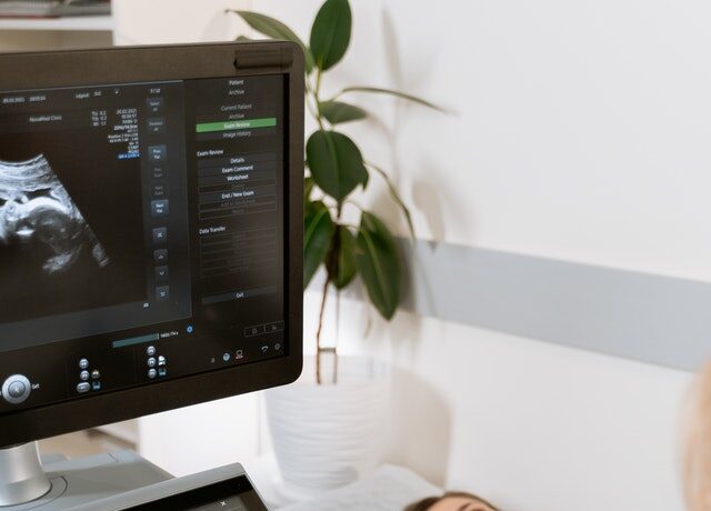
Many people don’t realize that ultrasound was first developed in the 1800s. However, it was not used in gynecology and prenatal care until 1958. The first 3D photos of a newborn were seen in 1986. 4D ultrasound, which allows you to view 3D pictures, was first introduced in the 1990s.
This generation’s prenatal care has been transformed by both traditional and 3D ultrasonography. Our moms and grandparents were given the care that was far different from what we get now. It’s critical to be able to spot issues and enhance the result for both mother and child. It’s also a thrilling experience to view your unborn child. If you want a great ultrasound that is conducted by a professional, we recommend that you check out the best portable ultrasound.

It is unsurprising that myths still persist, given all of this marvelous technology. This article should hopefully put any concerns to rest. This article should hopefully put any concerns to rest. Myth 1) The infant is harmed by ultrasounds. Ultrasounds do not seem to damage the fetus. There are potential hazards, such as heat generated, but all skilled sonographers follow safety standards that restrict the examination duration and keep the power settings as low as possible to collect the information required.
Myth 2) Ultrasound is the same as radiation. This is a complete lie. Ultrasound does not utilize radiation like X-Rays. Ultrasound employs high-frequency sound waves that are reflected when they meet the baby. The ultrasound machine’s computer then generates an image that appears on the screen.
Myth 3) In the first trimester, you should avoid having an ultrasound. This myth is most likely derived from the radiation myth, and it is untrue as stated above. An ultrasound scan does not seem to harm the infant. If a mother is experiencing pain or bleeding, many hospitals will utilize an early ultrasound to determine the baby’s viability. Couples who have lost children in pregnancy may desire to confirm their baby’s viability beforehand, which may provide a great deal of relief. In comparison to 2D ultrasounds, 3D ultrasounds employ greater sound waves. Both scans use the same frequency. The computer creates the 3D picture by layering many 2D images together.

Myth 4) Ultrasound allows you to know the gender of the baby 100% of the time. According to current data, an ultrasound can accurately determine the gender of a baby 97% of the time. In some instances, the baby’s location within the womb and the baby’s legs being crossed may compromise the precision of the evaluation.
Myth 5) Ultrasounds are only surface level examinations. If the examination is done on the abdomen, this is true. This occurs most often between 8 and 10 weeks of gestation and necessitates the use of a full bladder. A vaginal ultrasound may be suggested if it is only a few weeks into pregnancy and the baby is extremely little and low in the pelvis. Both the mother and the child are entirely safe. The probe is softly placed into the initial section of the vaginal canal and does not cause any discomfort. This method provides much sharper images of a little infant.


